CBC -The Nature of Things with David Suzuki - - The Nano Revolution: More than Human
This video apparently is only viewable in Canada. It is an excellent view of the future of nano technology at the "point of care" in medicine. It also has an excellent scenario of "post humans". It is worth watching just for the computer graphics effects on nanotechnology models.
Featured Post
Hacking Health in Hamilton Ontario - Let's hear that pitch!
What compelled me to register for a weekend Health Hackathon? Anyway, I could soon be up to my ears in it. A pubmed search on Health Hack...

Showing posts with label imaging. Show all posts
Showing posts with label imaging. Show all posts
Sunday, March 17, 2013
Saturday, August 4, 2012
Automatic Tape-collecting Lathe Ultramicrotome (ATLUM) device - In search of Immortality
I have always thought that one of the goals of ehealth was towards life extension, and this research article is indicative of the advances being made towards immortality, specifically - mind uploading - and a new word that I wonder might make head way in the English language lexicon - connectomics!
The strange neuroscience of immortality
July 30, 2012
[+]

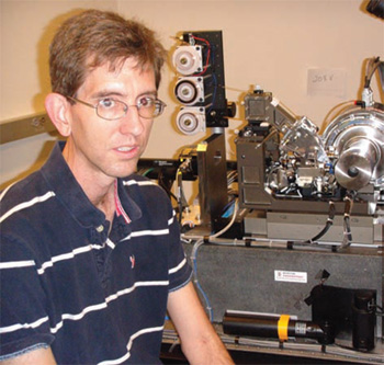
Kenneth Hayworth with his Automatic Tape-collecting Lathe Ultramicrotome (ATLUM) device (credit: Kenneth Hayworth)
Neuroscientist Kenneth Hayworth believes that he can live forever, the Chronicle of Higher Education reports. But first he has to die.
“The human race is on a beeline to mind uploading: We will preserve a brain, slice it up, simulate it on a computer, and hook it up to a robot body,” he says.
He wants that brain to be his brain. He wants his 100 billion neurons and more than 100 trillion synapses to be encased in a block of transparent, amber-colored resin — before he dies of natural causes.
The connectome grand theory
To understand why Hayworth wants to plastinate his own brain you have to understand his field — connectomics, a new branch of neuroscience. A connectome is a complete map of a brain’s neural circuitry. Hayworth looks at the growth of connectomics — especially advances in brain preservation, tissue imaging, and computer simulations of neural networks — and sees a cure for death.
Among some connectomics scholars, there is a grand theory: We are our connectomes. Our unique selves — the way we think, act, feel — is etched into the wiring of our brains. Unlike genomes, which never change, connectomes are forever being molded and remolded by life experience.
A human connectome would be the most complicated map the world has ever seen. Yet it could be a reality before the end of the century, if not sooner, thanks to new technologies that “automate the process of seeing smaller,” as Sebastian Seung puts it in his new book, Connectome: How The Brain’s Wiring Makes Us Who We Are.
Hayworth looks at the growth of connectomics — especially advances in brain preservation, tissue imaging, and computer simulations of neural networks — and sees something else: a cure for death. In a new paper in the International Journal of Machine Consciousness, he argues that mind uploading is an “enormous engineering challenge” but one that can be accomplished without “radically new science and technologies.”
Hayworth has founded the Brain Preservation Foundation, which offer a cash prize for the first individual or team to preserve the connectome of a large mammal. A dependable brain-preservation protocol is possible within five years, Hayworth says. “We might have a whole mouse brain preserved very soon.”
The foundation has published a Brain Preservation Bill of Rights on its Web site. ”It is our individual unalienable right to choose death, or to choose the possibility of further life for our memories or identity, as desired,” the document declares.
Hayworth’s brain-preservation and mind-uploading protocol
Before becoming “very sick or very old,” he’ll opt for an “early ‘retirement’ to the future,” he writes. There will be a send-off party with friends and family, followed by a trip to the hospital. After Hayworth is placed under anesthesia, a cocktail of toxic chemicals will be perfused through his still-functioning vascular system, fixing every protein and lipid in his brain into place, preventing decay, and killing him instantly.
Then he will be injected with heavy-metal staining solutions to make his cell membranes visible under a microscope. All of the water will then be drained from his brain and spinal cord, replaced by pure plastic resin.
Every neuron and synapse in his central nervous system will be protected down to the nanometer level, Hayworth says, “the most perfectly preserved fossil imaginable.”
Using a ultramicrotome (like one developed by Hayworth, with a grant by the McKnight Endowment Fund for Neuroscience), his plastic-embedded preserved brain will eventually be cut into strips, and then imaged in an electron microscope. The physical brain will be destroyed, but in its place will be a precise map of his connectome.
In 100 years or so, Hayworth says, scientists will be able to determine the function of each neuron and synapse and build a computer simulation of the mind. And because the plastination process will have preserved his spinal nerves, the computer-generated mind can be connected to a robot body.
“This isn’t cryonics, where maybe you have a .001 percent chance of surviving,” he said. “We’ve got a good scientific case for brain preservation and mind uploading.”
Thursday, May 3, 2012
Storing medical images on the cloud & the PHR
In my last post I mentioned novel ways of using Personal Health Records will be devised and here is just one example - storing and sharing medical images on the cloud. When I first read this is sounded like they had created a kind of mirror of a PACS server so the patients can access their medical image, and this they called a "Personal Health Record"! That would be novel, but I think I read the article too quickly. What is interesting here, is the idea that (finally) the patient "owns" the image (but not the cloud?) and can have it uploaded to a personal health record. The next great idea in phase III of this project will allow patients to consent to have the images used for clinical trial research - after de-identification. Besides organ donation there really should be some sort of data donation system when we die. Read it < here >
Sunday, April 29, 2012
Integrated Digital Pathology - no more microscopes?
At the Advances in Health Informatics Conference a few days ago, we saw several amazing presentations on digital pathology. Dr. Slyvia Asa talked about integrated digital pathology with telepathology, robotics, and streamlined processes for diagnoses to the point of care - all with a rapid turn around time. She quoted something by Sir William Osler, which I was able to find on the net for another talk she gave on pathology and informatics < here > and that was "As is your pathology, so goes your clinical care". When Osler was a young student he used to study bacteria from a reedy marsh not far from where I am typing this, and examined it with the greatest new technological marvel of that age - the microscope. Listening to Dr. Asa describe their integrated pathology labs at Toronto's University Health Network, it sounds like radiologists are exclusively using digital imaging to read pathological sample slices and make diagnosis. This was evident to me as well when I visited an anatomy lab in the Faculty of Health Sciences at McMaster, where the instructor showed us how digital imaging was being used more frequently than microscopes. He took a picture of a human cardio sample with his iPad, and then displayed the image on a big screen. He was able to zoom in with great detail on the pixels.
To quote from this article by Dr. Asa:
To quote from this article by Dr. Asa:
The future of pathology will be reports that are comprehensive clinical consultations that incorporate all of the imaging, biochemical, histologic, molecular, cytogenetic, and epigenetic data. Pathologists will not have two screens in front of them; most will have four. And they won’t want that microscope at their desk because it’s not good for their neck.
So what I see in the year 2020 as pathology is digital radiology, digital endoscopy, digital cardiology, digital genetics, all the relevant information on my four-screen computer in front of me, wherever I happen to be, always with quality assurance as an important part of it, and as the center of personalized medicine.
Subscribe to:
Posts (Atom)
-
Anxiety about coronavirus can increase the risk of infection — but exercise can help Stress about the coronavirus ...
-
I have tried several meditation apps and online meditation programs. I started out reluctantly because I didn't think the electronic...
-
FEB. 2, 2023 MEET THE SCIENTISTS WHO WANT TO MAKE MEDICAL DEVICES WORK FOR EVERYONE, FINALLY In the early months of the pandemic, Ashr...


